
If you’ve been told you need an angiogram, you might feel a mix of anxiety and curiosity.
An angiogram, also known as cardiac catheterization, is a common medical procedure.
In fact, it’s performed more than 1,000,000 times each year in the United States alone.
With such a high number of procedures, you might wonder about the risks involved, especially the risk of death.
What Are the Risks?
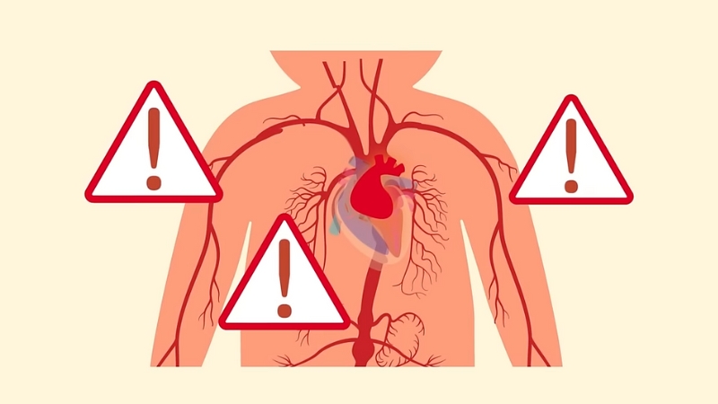
Despite its routine nature, angiogram carries risks of death. The good news is that the risk of major complications is relatively low. Based on NCBI claims, here are some key statistics:
- Major Complications: Less than 1% during diagnostic procedures. These include serious events like heart attacks, strokes, or major bleeding, but they are uncommon due to careful patient selection and advanced procedural techniques.
- Risk of Death: Approximately 0.05% for diagnostic procedures, meaning about 1 in 2 (6.45 cm²),000 patients might face this risk. While any risk of death can be alarming, it is important to consider this in the context of the procedure’s benefits and the life-saving information it can provide.
- Retroperitoneal Bleeding: Less than 0.2% incidence. This rare but serious complication involves bleeding into the space behind the abdominal cavity and can require urgent medical attention.
- Radial Artery Occlusion: About 5% of cases use the transradial approach. While this complication can occur, it is often asymptomatic and can be managed with appropriate care and follow-up.
- Stroke: Risk ranges from 0.05% to 0.1% in diagnostic procedures and 0.18% to 0.4% in interventional procedures. Strokes are a serious concern, but their risk is minimized through careful procedural techniques and patient monitoring.
- Contrast-Induced Acute Kidney Injury: Occurs in 7.1% of patients undergoing coronary interventions. This risk is higher in patients with pre-existing kidney problems and requires careful management of hydration and medication.
- Allergic Reactions: Up to 1% of patients may react to contrast agents used during the procedure. Reactions can range from mild to severe, and pre-procedure screening helps identify patients at risk.
The Basics of the Procedure
在 Instagram 查看这篇帖子
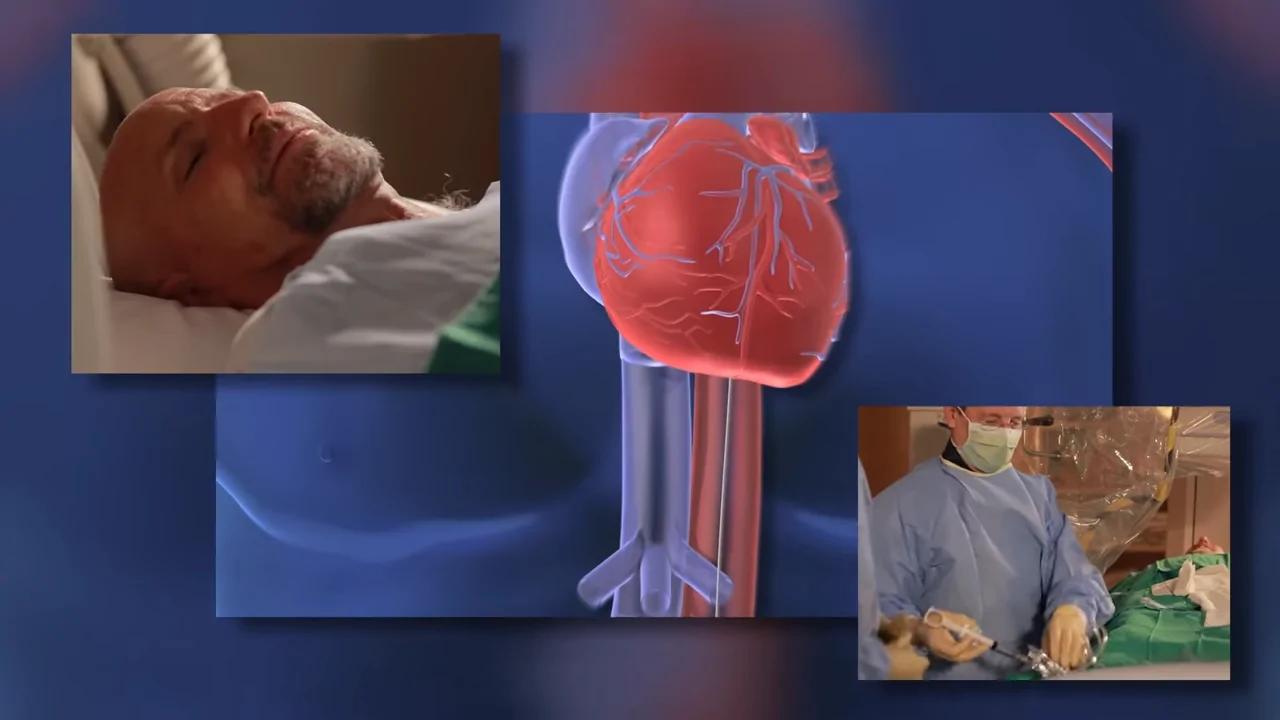
Cardiac catheterization is a medical procedure where doctors insert a thin, flexible tube called a catheter into a blood vessel.
This catheter is then guided to the heart to diagnose and sometimes treat various heart conditions.
The process might sound complex, but it has become a routine procedure thanks to advancements in medical technology and techniques.
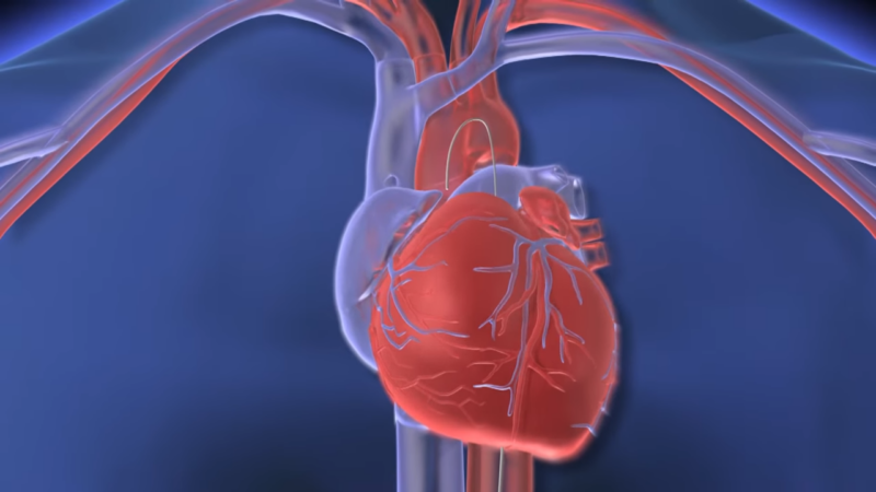
The goal of the procedure is to provide detailed information about the heart’s function and identify any issues that may require treatment.
It can also be used to perform certain treatments, such as angioplasty, to open up blocked arteries.
Why Is Cardiac Catheterization Done?
Doctors recommend cardiac catheterization for several reasons:
Coronary Artery Disease
To check for blockages in the heart’s arteries, a common complication in heart disease. Blockages in these arteries can reduce blood flow to the heart muscle, leading to chest pain (angina), shortness of breath, or even heart attacks.
By identifying these blockages, doctors can decide on the best treatment, such as medications, angioplasty, or surgery.
Measuring Hemodynamics
To assess blood flow and pressure in the heart. This is important for diagnosing and managing various heart conditions, such as heart failure or pulmonary hypertension.
Accurate blood pressure (BP) measurement is essential for the diagnosis and management of hypertension. In clinical practice, BP is estimated using noninvasive methods with significant variability of application of guidelines in clinical practice, impacting the accuracy and certainty of BP measurements. Martha Gulati
Evaluating Left Ventricular Function
To see how well the heart’s left ventricle pumps blood. The left ventricle is the main pumping chamber of the heart, and its function is crucial for maintaining adequate circulation.
Assessing left ventricular function helps in diagnosing and managing conditions like heart failure, cardiomyopathy, and valve diseases.
Cardiac Arrhythmias
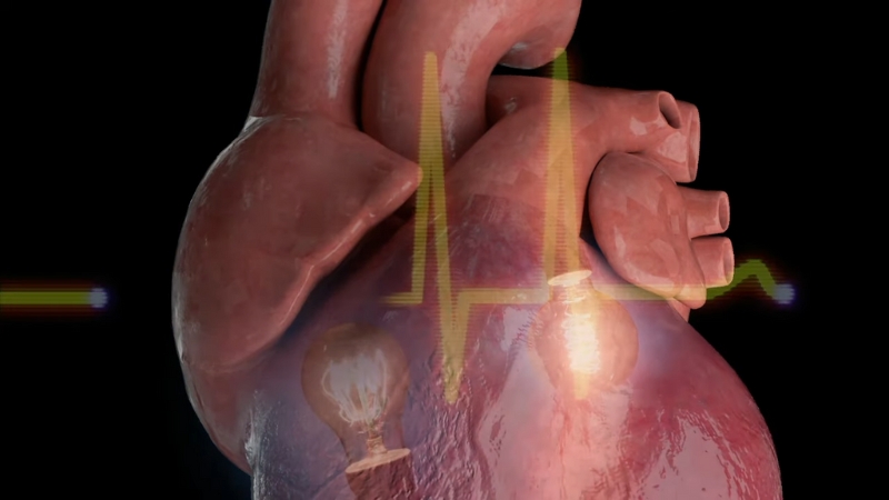
To evaluate and sometimes treat irregular heartbeats. Abnormal heart rhythms can cause symptoms like palpitations, dizziness, or fainting and can lead to serious complications if left untreated. During cardiac catheterization, doctors can map the electrical activity of the heart and, in some cases, perform procedures to correct the arrhythmias, such as ablation.
Valvular Heart Disease
To assess and plan treatment for heart valve problems.
Heart valves ensure the proper flow of blood through the heart’s chambers, and issues with these valves can lead to symptoms like shortness of breath, fatigue, and heart failure.
Cardiac catheterization provides detailed information about valve function, helping doctors plan treatments such as valve repair or replacement.
Pericardial and Myocardial Diseases
To examine the heart’s outer and muscle layers.
Conditions affecting the pericardium (the sac surrounding the heart) or the myocardium (the heart muscle) can cause chest pain, fluid buildup, and impaired heart function.
Pericardial effusion is the abnormal accumulation of fluid in the pericardial sac, often caused by pericarditis or other conditions such as hypothyroidism or heart rupture. It can lead to cardiac tamponade, which obstructs the heart and can result in low blood pressure and, in extreme cases, cardiac arrest. Ary L. Goldberger MD
Cardiac catheterization can help diagnose these conditions and guide appropriate treatments.
Congenital Heart Disease
To evaluate heart defects present from birth. Congenital heart defects can impact the structure and function of the heart and may require treatment during infancy, childhood, or adulthood.
Cardiac catheterization provides detailed images and measurements that are essential for planning surgeries or other interventions according to an NCBI study.
Heart Failure
To help in managing and treating heart failure.
Heart failure occurs when the heart is unable to pump enough blood to meet the body’s needs.
By assessing heart function and pressures, cardiac catheterization helps in tailoring treatments to improve symptoms and quality of life for heart failure patients.
Although clinical tools for the diagnosis and treatment of heart failure (HF) have been developed, mortality and morbidity rates for HF remain steady, and optimal medical therapy rates remain low. This indicates that there is a gap between clinical practice and guidelines and highlights the importance of quality assessment of HF care. This review article examines HF treatment patterns and treatment adherence in real-world practice, identifies clinical gaps, suggests ways to improve the quality of care for HF, and finally aims to improves care and clinical outcomes for patients with HF. Se-Eun Kim
Preparing for the Procedure
Preparation involves a thorough patient history and physical examination.
Doctors also require specific lab work, such as a complete blood count (CBC), basic metabolic panel (BMP), prothrombin time, electrocardiogram (ECG), and chest X-ray.
They pay special attention to any allergies and manage chronic kidney disease to prevent complications.
This careful preparation ensures that the patient is in the best possible condition for the procedure and that any potential risks are minimized. It also provides vital information that helps the medical team plan the procedure and anticipate any challenges that may arise.
Which Techniques Are Used?
The two main access points for catheter insertion are the femoral artery in the groin and the radial artery in the wrist.
- Femoral Access: Doctors use anatomical landmarks or imaging techniques like fluoroscopy or ultrasound to guide the catheter. This traditional approach provides a direct path to the heart but can have a higher risk of complications such as bleeding or vascular damage. The choice of femoral access may depend on the patient’s anatomy, the type of procedure, and the operator’s preference and experience.
- Radial Access: This approach is preferred because it has fewer complications. Tests like the Allen or Bar Beau test ensure adequate blood flow before access. Radial access allows patients to move around more quickly after the procedure and typically results in a shorter hospital stay. It also reduces the risk of serious bleeding complications, making it a safer option for many patients.
How to Minimize Risks
Advancements in medical technology and techniques have significantly reduced the risks of death associated with angiograms. For instance:
Transradial Approach
This method has been shown to reduce mortality, major bleeding, and other complications compared to the transfemoral approach. It also allows for quicker recovery and shorter hospital stays, making it a preferred option for many patients and physicians.
Ultrasound Guidance
Ultrasound to guide vascular access is one of the most elegant innovations in modern medicine.
Almost no cost/downside. Not sure it requires RCT
*Different from ICE/TEE, which entail lots of extra cost and some risk. https://t.co/OFt7iXyhsc
— John Mandrola, MD (@drjohnm) September 9, 2024
Using ultrasound to guide femoral access reduces the risk of complications by 49%. This technology helps ensure accurate placement of the catheter and minimizes the chances of vascular damage or bleeding.
Complications in Detail
- Local Vascular Complications: These include hematoma, bleeding into the retroperitoneal space, pseudoaneurysm, arteriovenous fistula, dissection, thrombosis, and embolism. These complications can cause pain, and swelling, and, in severe cases, require additional medical intervention.
- Transradial Access Complications: These might include radial artery occlusion, spasm, and, rarely, perforation. While these issues are less common than with femoral access, they can still occur and require careful management to avoid long-term problems.
- Major Complications: These include death, myocardial infarction (heart attack), stroke, dissection, or perforation of major blood vessels, atheroembolism (debris causing blockage in blood vessels), allergic reactions, acute kidney failure, infection, radiation injury, and arrhythmias (irregular heartbeats). Each of these complications, although rare, can have serious consequences and requires prompt recognition and treatment by the medical team.
Methodology
- Research: Gathered reliable data from trusted medical sources like NCBI and clinical studies on angiograms.
- Organized Structure: Divided content into clear sections covering the procedure, risks, and benefits for easy reading.
- Data Integration: Included key statistics on risks and outcomes to provide a fact-based overview.
- Simplified Language: Translated complex medical terms into accessible language.
- Balanced Perspective: Highlighted both risks and benefits to offer a well-rounded view.
- Relevant Examples: Used examples to clarify risks and the impact of medical advancements.
In Summary
Cardiac catheterization, or an angiogram, is an essential procedure in modern cardiology.
While the risk of death exists, it remains very low, especially with the continuous improvements in medical practices.
Always consult with your healthcare provider to understand how these risks apply to your specific situation and what steps are taken to ensure your safety.